






















RADIOLOGY
quantusTX uses Artificial Intelligence to analyze chest X-rays and automatically detect atelectasis, consolidation, pneumothorax and pleural effusion.
AI-based detection of chest pathologies in minutes.

DESCRIPTION
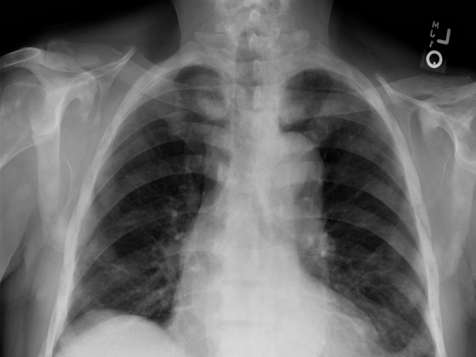
quantusTX is an artificial intelligence-based test that automatically detects chest pathologies from chest X-rays.
quantusTX is a software medical device (MDSW) designed for use by radiologists and other healthcare professionals to support the evaluation of chest X-rays. The system analyzes the image and provides a risk estimate (expressed as a percentage) for atelectasis, consolidation, pneumothorax and pleural effusion. The device provides medical information to support decision-making in the management of patients at risk of a chest pathology.
The device is designed to be used as a diagnostic support tool; the results report provides the medical specialist with objective information to support clinical decision-making.
USE CASES

CU-TX-01: Screening in Primary Care
In primary care, the physician orders a chest X-ray when a chest pathology is suspected. quantusTX automatically analyzes the image and generates a report with the risk estimate for atelectasis, consolidation, pneumothorax and pleural effusion to support clinical decision-making. The physician uses the report to support clinical decisions, which speeds up patient screening, optimizes workflow, and reduces costs.
CU-TX-02: Support for detecting urgent findings in the emergency department
For a patient with acute respiratory or chest symptoms, a chest X-ray is taken and immediately analyzed by quantusTX, flagging the possible presence of findings requiring urgent action, such as pneumothorax or significant pleural effusion, while awaiting the definitive radiological reading.
CU-TX-03: Second reading and prioritization in radiology departments
In radiology departments with a high volume of chest X-rays pending reporting, quantusTX systematically analyzes them and provides a risk estimate for each one, helping to order the radiologist’s worklist and prioritize the reading of studies with the highest likelihood of relevant findings.

CLINICAL EVIDENCE
Automatic detection of pathologies in chest X-rays (quantusTX).
The study used images from public databases: the Valencian Region Medical Image Bank (BIMCV) and the Stanford Machine Learning Group. The development of the tool consists of the following steps:
The results obtained for each pathology are as follows:
| Pathology | Sensitivity | Specificity | PPV * | NPV * |
|---|---|---|---|---|
| Atelectasis | 86.6% | 71.5% | 75.1% | 84.4% |
| Consolidation | 89.1% | 80.6% | 81.2% | 88.8% |
| Pneumothorax | 87.2% | 80.3% | 81.4% | 86.4% |
| Pleural Effusion | 92.4% | 88.9% | 90.2% | 91.3% |
* PPV and NPV: Positive Predictive Value and Negative Predictive Value

SOLUTION INTEGRATION
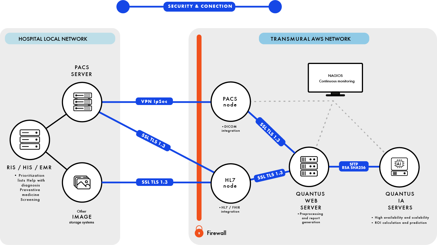
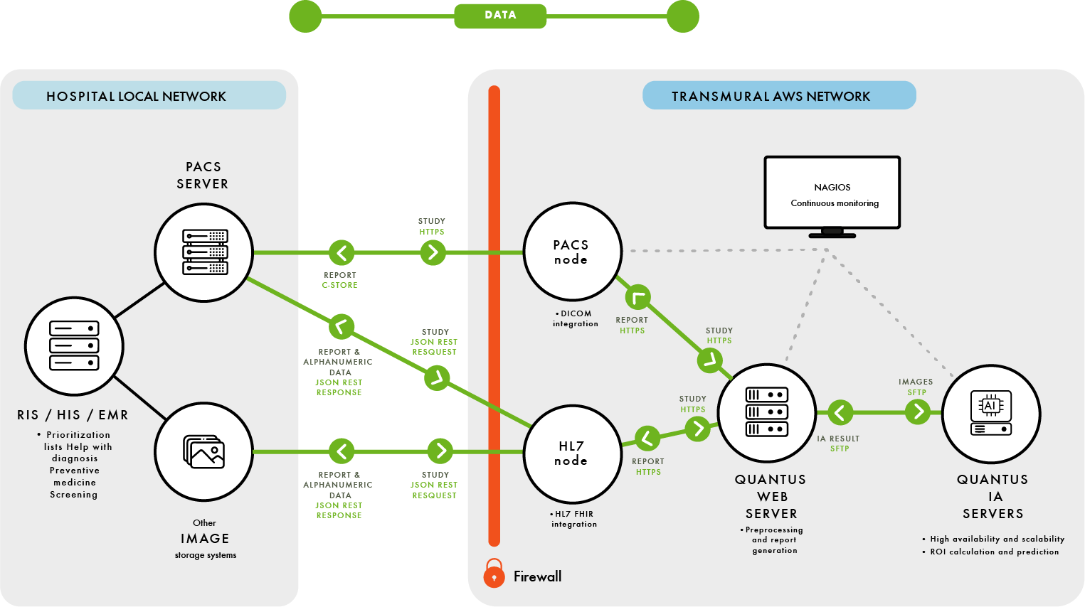
- Our products allow integration with client systems through the DICOM protocol and the HL7 FHIR interface
- We ensure data privacy in SaaS installations by establishing IPsec VPNs with our clients.
- All communications outside the client environment are secured with SSL TLS 1.3

LICENSING BY NUMBER OF TESTS
LICENSING BY ANALYSES PERFORMED
CUSTOM OFFERS


