






















DERMATOLOGY
.
Assesses the risk of malignancy in skin lesions.
From dermoscopic images or photos taken with a smartphone, quantusSKIN lets you classify the malignancy risk for different types of melanoma.

DESCRIPTION
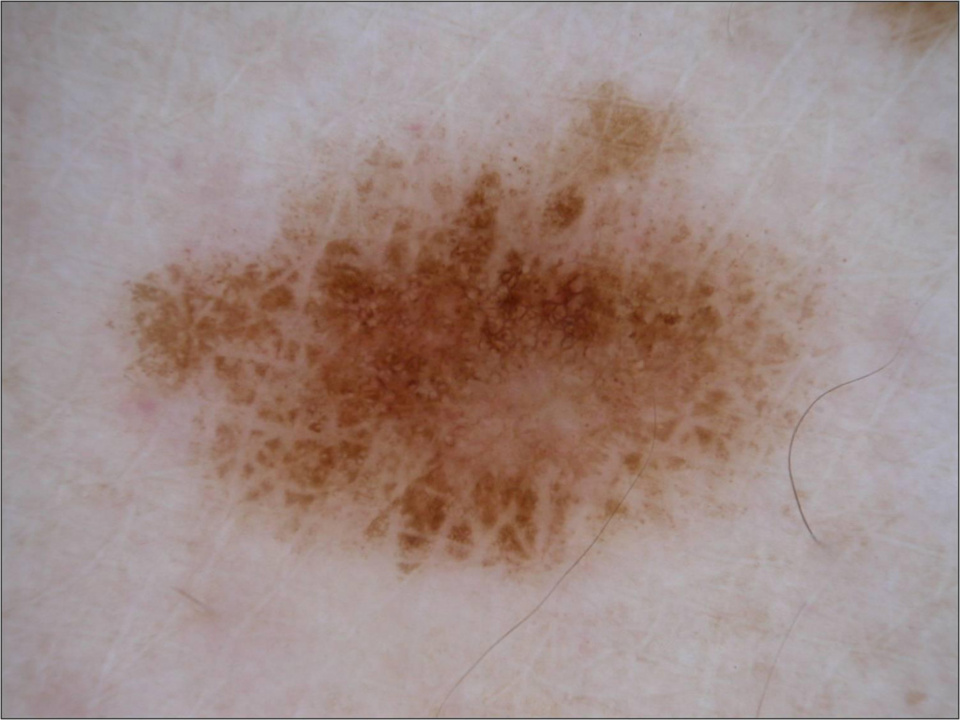
quantusSKIN estimates the probability that a skin lesion is benign or malignant from dermoscopic images.
It is a software medical device (MDSW) based on artificial intelligence (AI), designed for use by dermatologists and other trained healthcare professionals, which analyzes dermoscopic images and estimates the probability that the image represents a benign or malignant lesion.
The device provides medical information to support decision-making related to the evaluation and management of patients with suspicious skin lesions.
USE CASES

CU-SKIN-01: Dermatological Screening from Primary Care and Family Medicine
In primary care, quantusSKIN analyzes images of skin lesions obtained with a smartphone or dermatoscope and provides a malignancy risk classification within minutes, helping the healthcare professional prioritize referral to the specialist.
CU-SKIN-02: Teledermatology and Remote Consultation
quantusSKIN has a teledermatology system that allows skin lesion images to be sent via WhatsApp. These are automatically analyzed to generate a preliminary report that supports the dermatologist, enabling a faster, more objective and efficient teleconsultation.
CU-SKIN-03: Support Tool for Early Melanoma Detection Campaigns
In population screening campaigns, quantusSKIN analyzes skin lesion images obtained by healthcare professionals in real time, classifying their risk level and immediately identifying cases that require priority dermatological evaluation.
CU-SKIN-04: Diagnostic Support in Hospital Dermatology Units
In dermatology departments with high patient loads, quantusSKIN automatically pre-classifies the risk of skin lesions photographed during the consultation, allowing the dermatologist to prioritize the most suspicious cases and document their clinical decision alongside the algorithm’s result.

CLINICAL EVIDENCE
Tool for the detection of malignant skin lesions from dermatological images or photographs (qSKIN).
More than 50,000 dermoscopic images available online from the International Skin Imaging Collaboration (ISIC) Archive were used, acquired at different centers worldwide and labeled by recognized dermatologists with different types of benign and malignant skin lesions.
The development of the tool consists of several steps:
The following results were obtained:
| Accuracy | Sensitivity | Specificity | PPV * | NPV * |
|---|---|---|---|---|
| 85.9% | 89.6% | 85.2% | 52.6% | 97.8% |
* PPV and NPV: Positive Predictive Value and Negative Predictive Value

SOLUTION INTEGRATION
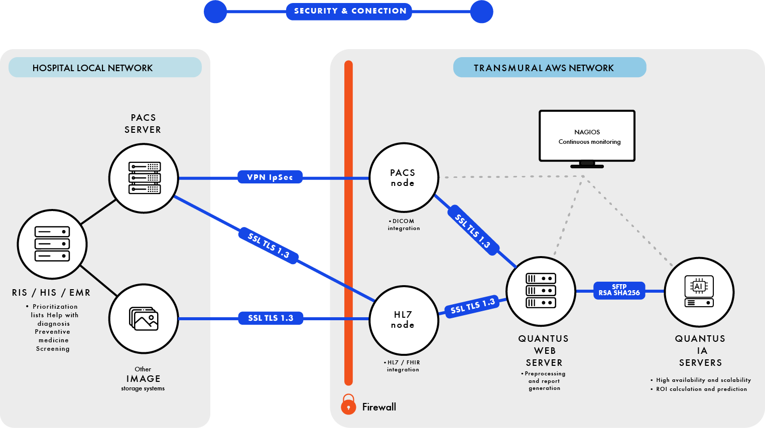
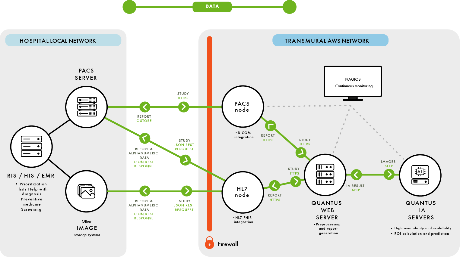
- Our products allow integration with client systems through the DICOM protocol and the HL7 FHIR interface
- We ensure data privacy in SaaS installations by establishing IPsec VPNs with our clients.
- All communications outside the client environment are secured with SSL TLS 1.3

LICENSING BY NUMBER OF TESTS
LICENSING BY ANALYSES PERFORMED
CUSTOM OFFERS


